
 |
|
 |
|
 |
|
|
|
 |
|
|
|
 |
|
|
|
 |
|
|
|
 |
|
|
|
 |
|
|
|
 |
|
|
|
 |
|
|
|
 |
|
|
|
 |
|
|
|
 |
|
|
X-ray Fluorescence Imaging For the April 2004 workshop three different scientists suggested independently that we employ a technique called X-ray Fluorescence Imaging. This technique would involve exciting the Palimpsest with relatively high energy, short wavelength photons (X-rays). Normal X-ray images are taken in transmission: that is, a reactive plate is placed behind a sample, and a negative image of an object is created on the plate. This is what happens when you go to the dentist. However, this transmission technique would not work for the Palimpsest because the image contrast from the traces of ink would be much too small. So they proposed to use a different way to enhance the contrast. Some of the X-rays that do not pass through the leaves excite individual atoms in the palimpsest, which would then generate their own secondary X-rays. The crucial thing about these so called fluorescence X-rays is the fact that different elements generate X-rays with different wavelengths. A fluorescence X-ray sent out from an iron atom is different from an X-ray sent out by a calcium atom, or an atom of gold. These fluorescence X-rays could then be detected with a device that can recognize these different X-rays. The idea is simple: by mapping these X-rays, we could create �element maps� of individual pages of the Palimpsest. The imaging scientists who worked on this aspect of the project were Gene Hall, Professor of Chemistry at Rutgers University, Uwe Bergmann, Staff Scientists at the Stanford Synchrotron Radiation Laboratory, and Bob Morton of the Children of the Middle Waters.
Initial experiments were conducted by Gene Hall at Rutgers University, using an X-ray microfluorescence system provided by the EDAX Corporation — an EDAX Eagle Probe — in his lab. He presented his initial results in April 2004. If you would like to see his presentation, click here. Following the conference it was decided to do further imaging with an EDAX Eagle probe. The EDAX firm, a subsidiary of Ametek, were extremely generous in providing their facilities in New Jersey for five days of intensive research by Bob Morton and Gene Hall.
They were greatly helped in their efforts by Tara Nylese and Bruce Scruggs of EDAX inc.
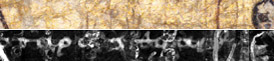
There were notable results from this imaging effort.
These images were exciting, but the small section of Archimedes text that we recovered from the forged page took 10 hours to generate! It was clear that we would need more intense X-rays, and for this we needed a more powerful instrument. It was for this reason that we turned to Uwe Bergmann, and to the Stanford Synchrotron Radiation Laboratory (SSRL).
If you would like to see Uwe Bergmann�s proposal for imaging the Palimpsest, click here. The important point about synchrotron radiation is that it is much more powerful than the radiation that a normal X-ray tube can provide, and it is also much more tunable to the specific wavelengths that we wanted. It therefore had the potential to produce similar results to those of the EDAX Eagle Probe, but much more quickly. The potential danger of using synchrotron radiation is that these high energy X-rays have the potential to damage the parchment. Therefore careful tests were carried out by Gregory Young of the Canadian Conservation Institute, to see how parchment reacted to exposure to X-rays, and we were guided by these results at Stanford. If you would like to see this report, click here.
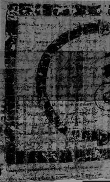
The results can be seen below. We retrieved a whole column of Archimedes text from the same forged page of the Palimpsest in about 24 hours. If you would like to see a summary of these efforts in more detail, click here.
|
|||||||||||||||||||||||||||||||||||||||||||||||||||||||
|
Top of page
|
|||||||||||||||||||||||||||||||||||||||||||||||||||||||
 Tara Nylese


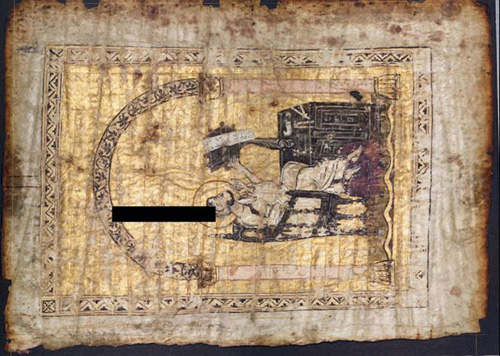
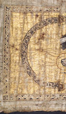
 This image is of one of the pages in the Archimedes Palimpsest containing a forgery. A small section was imaged, and the resulting iron map produced a section of the Archimedes �Equilibrium of Planes�, which our scholars could decipher.
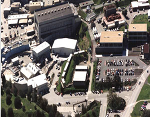
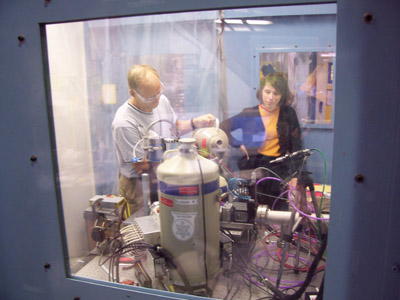 For this experiment we used Beamline 6 at the SSRL, which is encased within a lead lined hutch.
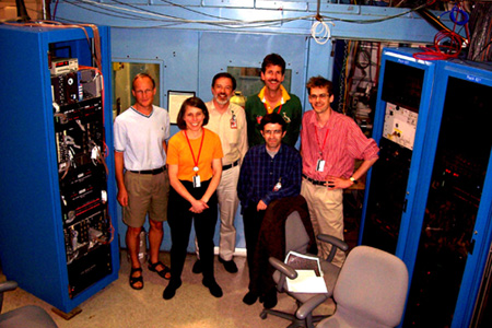
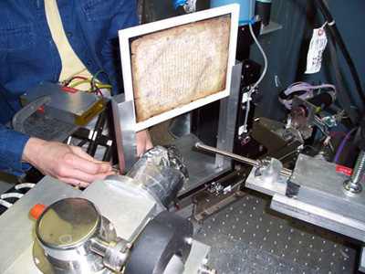 A leaf of the Archimedes Palimpsest was mounted vertically on an XY stage, and placed before the beam of high energy photons. A detector, tuned to pick up X-rays of the wavelength on Iron was placed at 90 degrees to the beam. Mike Toth, Will Noel, Abigail Quandt, Uwe Bergmann, Martin George and others worked for six days on these experiments.